Requirement for microscope:
A, binoculars, double light sources (electric or nature light) the better,
B, Designed with a mechanical mobile platform,
C, Eue len 16x Objeet: low10x, high40x
2, Sebaceous dissolvent: limpid vegetable oil (bean oil,rape-seed oil,sesame oil and salad oil bought from market are all valid, don’tuse liquid paraffin, glycerin, shampoo oil, normal saline, etc,)
3, Little bottle for holding oil and a pipette,
4, Latex glove for thumb and second finger,
5, Absorbent cotton ball,
6, Loading glass,
7,Taking mite apparatus, sebaceous quantitative apparatus (designedparticularly and pattern enclosed) acne needle,
8, Sterile solution:3%--5%liquor cresolis saponatus (Lysol)
(2), Get specimen for inspecting Demodex
The method for getting sebaceous:The sebaceous gland of skin hair follicle of human face is the best place for the growth and breed of human larva form acarid, So we have to get sebaceous first, if we want toinspect the Demodex
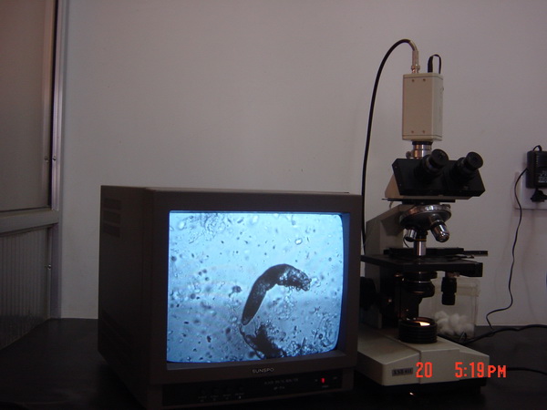
Method: First, let your thumb and second finger wear latex glove, then, drop three guttae vegetable oil on the glass respectively, The doctor and patient sit down face to face, the doctor, using right hand thumb and second finger worn glove, squeeze the nose top and nose sulcus of the patient, when the sebaceous had squeezed out, you can get the sebaceous off with the taking mite apparatus, and put it in the oil gutta, which is on the glass center, and dissolve it Then, using your left thumb, push the patient`s frontal skin up, meanwhile let the patient look down, then, with your right hand hold the taking mite apparatus, its point side back to inside and face down, shave the sebaceous off,and put it in the other oil gutta on the loading glass, dissolve it, then, let patient gnash his lower lip, With right hand, you can hold taking mite appartus, to shave the patien’s submandibularis skin, from outside to inside and downwards, when you get the sebaceous, put it in the third oil gutta on theloading glass, dissolve it Or,you can also use the round head part of the acne needle to press and shave the place described above, to get the sebaceous and put it in the oil gutta, and dissolve it then, put the taking mite apparatus in Lsol to sterilize it, prepared for use Get off the glove, put the loading glass on the platform of the microscope examining it with low power first, modify the focus and light,as tosee, the mite and masses in oil clwarly, to find mite in oil guttae, include living and dead adult worm, immobile worm eggs, nymph and remain bodies, by draw net style , and differentiate long form mite from short form mite , All the mites showed the form described above being inspeeted should count as infectton level, if not clearly. You may transfer to high power microscope, If you can ‘t find out the mtie in sebaceous that you had gotten at first time (naybe the method you used qas not propetly )you can get the sebaceous again with the same method to examine.
Enclose:The morphology for all period of human larva form acarid,
An experienced doctor may complete an inspecting mite diagnosis in 5—10minutes in common.
Notice: When adult mite get off from human body, and was put in vegetable oil, the long and short fofm mite both can nove about , in temperature 25—36℃,some can live 36—58 hours, its most dynamic temperature is 28℃--33℃.Under 25℃,living mite will stop its motion, So,in winter, when room trmperature under 25℃,you have to hold the glass, which loading the dissolved sebaceous , in palm to worm it for three minutes, examining it under microscope at once. till then,you can see the adult mite motion, Worm eggs, larvae, nymph and dead mite can not move , If you observe nymph under high power microscope carefully, you may see its mouth moving
If you want to do medical research, you may put the gotten sebaceous in the sunken groove (0.5mm)of the sebaceous quantitative apparatus, press it even, then take off thes certain volume sebaceous by the point taking mite apparatus, and put it in the oil gutta on the loading glass, dissolve it, then observe it under the microscope carefully, count the number of the mites, write down the lomg and short form mites number respectinely and the palce they are from, include worm eggs, larvae and remain bodies, as to compare with the number inspected in same place the next time.
With the method described above, when squeezing sebaceous, some patients feel uncomfortable, and you may see through the movement of the mites, the oid method, patch a 1cm transparent adhesive tape on the patient’s nose alar last night , and take it off in the morning the second day, and put it on the glass, then, observe it undeeer microscope, Although, with this method, patient have not any pain described above, but was time –consuming and was not accurate , Sonmetimes, patient will scrub the tape off in sleeping , So,you hane to do it again , By this method , you can see the dead adult mites only, Generally, you can’t see the worm egg, larva or nymph , you can’t do science count also, The oid method hhad been ahandoned long ago.
Chinese specialist of preventing and treating human larva form acaridiasis:ZhaoZhong-zhou